High Arches
(Pes Cavus) at
Foot Foundation
High Arches (Pes Cavus) describes an excessively high arch, which reduces shock absorption and overloads the heel and forefoot. Patients often experience arch pain, instability, or recurrent sprains.
At Foot Foundation, we provide cavus-specific orthotics, footwear guidance, strengthening, and rehabilitation programs to reduce pain, improve stability, and protect against long-term complications.
What are High Arches (Pes Cavus)?
Pes cavus describes an excessively elevated medial longitudinal arch. This can be flexible or rigid, and may be driven by the forefoot (e.g., plantarflexed first ray) or the hindfoot (often with hindfoot varus). The altered foot shape concentrates pressure under the heel and forefoot, reduces shock absorption, and predisposes to lateral ankle instability, metatarsalgia, stress fractures, and peroneal tendinopathy.
In some patients, pes cavus is idiopathic (no clear cause). In others, it is associated with neuromuscular conditions (e.g., Charcot–Marie–Tooth disease), previous trauma, or long-standing biomechanical adaptation. Determining the driver of the deformity (forefoot- vs hindfoot-driven) is essential, as it informs targeted treatment.
At Foot Foundation, we perform a comprehensive biomechanical and neurological screen and design a precise management plan to redistribute load, improve stability, and reduce pain.
Causes & Risk Factors
Structural/biomechanical factors
Forefoot-driven cavus (plantarflexed first ray, forefoot valgus/varus)
Hindfoot-driven cavus (hindfoot varus, calcaneal inversion)
Rigid cavus increasing lateral column loading
Neuromuscular conditions (variable severity)
Charcot–Marie–Tooth and other hereditary neuropathies
Post-stroke or spinal conditions causing muscle imbalance
Overload & activity
High-impact sports, hill running, abrupt training changes
History of injury
Recurrent lateral ankle sprains, peroneal tendon injury
Footwear factors
Minimal cushioning or narrow toe boxes exacerbating pressure
Family history / genetics
Familial foot structure patterns and neuromuscular predisposition
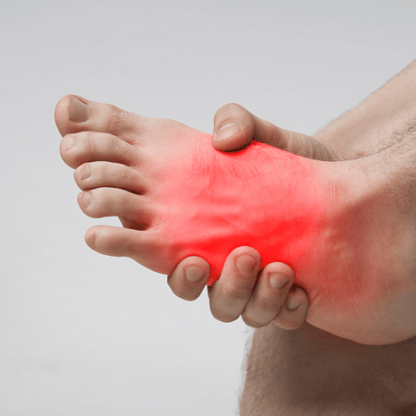
Symptoms
Localised forefoot pain (metatarsalgia, sesamoid pain), callus build-up under metatarsal heads
Heel pain from concentrated impact forces
Recurrent ankle sprains or “rolling out” (hindfoot varus/lateral overload)
Peroneal tendinopathy (pain along the outside of the ankle)
Claw toes or hammertoes from intrinsic muscle imbalance
Foot fatigue, difficulty on uneven ground, poor shock absorption
In long-standing or severe cases: lateral column overload, stress reactions/fractures
Diagnosis
At Foot Foundation, assessment focuses on identifying the mechanical driver and secondary pathology:
Weight-bearing exam & gait analysis (frontal/transverse plane alignment, stride mechanics)
Coleman block test to distinguish forefoot-driven vs hindfoot-driven cavus
Range-of-motion & strength testing (peroneals, tibialis posterior, calf)
Pressure distribution assessment (forefoot/heel focal loading)
Neurological screen (sensation, reflexes, muscle power) where indicated
Imaging when needed:
X-ray (alignment, 1st ray position, hindfoot varus)
Ultrasound/MRI for peroneal tendons, stress injury, sesamoids
Neuro referral if neuromuscular disease suspected
Treatment at Foot Foundation
Goal: redistribute load, improve stability, protect soft tissues, and address the deformity driver.
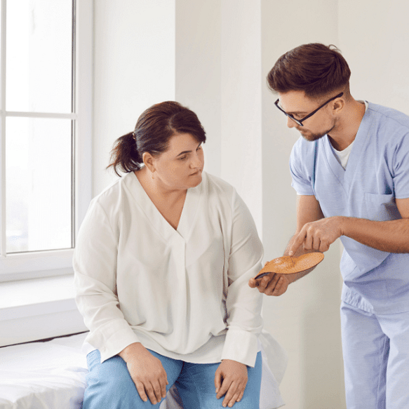
Custom Orthotics (cavus-specific design)
Forefoot-driven: first-ray cut-out / valgus forefoot posting to let the first ray drop
Hindfoot-driven: lateral wedging/valgus rearfoot posting to reduce varus and lateral overload
Cushioned top covers to reduce impact under heel/forefoot; metatarsal pads to offload
Intrinsic/extrinsic posting guided by gait and Coleman block findings
Footwear Prescription
Stable, cushioned trainers with adequate midsole thickness
Rocker soles to reduce forefoot loading in rigid cavus
Wider toe box to reduce claw-toe pressure; avoid overly minimal shoes
Rehabilitation
Peroneal strengthening and lateral stability work
Calf flexibility (gastrocnemius/soleus) and ankle mobility
Proprioception/balance retraining for lateral instability
Intrinsic foot muscle conditioning
Adjunct Therapies
Strapping/bracing for sport or unstable terrain
Shockwave therapy for chronic peroneal or insertional tendon pain
Foot Mobilisation Therapy to improve joint mechanics when appropriate
Referral Pathways
Neurology when neuromuscular disease suspected
Orthopaedics for rigid, painful deformity unresponsive to conservative care (e.g., osteotomy, tendon transfer, soft-tissue balancing)
High Arches – FAQs
Pes cavus is a foot type with an excessively high arch, often combined with hindfoot varus and forefoot imbalance. It reduces shock absorption and concentrates pressure under the heel and forefoot, increasing injury risk.
Causes include structural/biomechanical patterns, prior injury, and neuromuscular conditions (e.g., Charcot–Marie–Tooth). Some cases are idiopathic (no clear cause). Family history and genetics can play a role.
Common issues include metatarsalgia, sesamoid pain, peroneal tendinopathy, recurrent ankle sprains, claw toes, calluses, and stress fractures from focal loading.
We use weight-bearing assessment and the Coleman block test. If hindfoot varus corrects when the first ray is unloaded, the deformity is forefoot-driven; if not, it is hindfoot-driven—guiding orthotic design and posting.
Orthotics don’t change bone shape, but they do redistribute load, improve alignment, and reduce pain. Cavus-specific orthoses (e.g., first-ray cut-out, lateral posting, cushioning) are highly effective for function and comfort.
Choose cushioned, stable footwear with adequate midsole thickness and a roomy toe box. Rocker soles can reduce forefoot load in rigid cavus. Avoid overly minimal or stiff shoes that increase pressure and instability.
Yes. Peroneal strengthening, proprioception/balance training, and calf flexibility improve lateral stability and reduce overload. Intrinsic foot strengthening can aid toe function and pressure distribution.
Hindfoot varus in cavus feet biases weight to the lateral column, predisposing to lateral ankle sprains. Lateral posting, balance work, and peroneal strengthening address this risk.
Yes. Reduced shock absorption and altered alignment can transmit forces up the kinetic chain, contributing to shin pain, knee symptoms, hip/back discomfort.
Some rigid patterns remain stable; others progress—especially with underlying neuromuscular drivers or recurrent injury. Early, targeted management reduces complications and maintains function.
Only when conservative care fails and deformity is rigid, painful, or progressive. Procedures may include osteotomy, tendon transfer, or soft-tissue balancing. We coordinate assessment and referral when appropriate.
If you have forefoot or heel pain, recurrent ankle sprains, or callus build-up, or if your foot feels unstable on uneven ground, seek assessment. Early intervention prevents secondary problems.
Why Choose Foot Foundation?
Foot Foundation delivers specialist-level assessment and treatment for pes cavus, integrating podiatry and physiotherapy. We use cavus-specific orthotics, footwear optimisation, stability rehabilitation, and adjunct therapies to reduce pain and restore confidence in movement. Neurological and orthopaedic pathways are coordinated when needed.
With clinics in Rosedale, Takapuna, Remuera, Botany, Hamilton, and Tauranga, expert care is available across New Zealand.