Stress Fracture Treatment at Foot Foundation
Stress fractures are tiny cracks from cumulative overload—common in the tibia, fibula, and foot bones. Pain localises, worsens with impact, and can persist at rest if ignored.
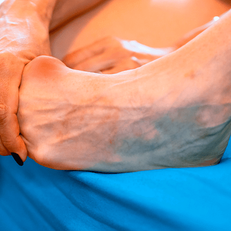
At Foot Foundation, we prioritise early recognition, off-loading and protective footwear/boots, custom orthotics, and progressive rehab with clear return-to-sport milestones.
What are Stress Fractures?
A stress fracture is a small crack in a bone caused by repetitive loading that exceeds the bone’s ability to repair itself. Unlike acute fractures from trauma, stress fractures develop gradually due to overuse and cumulative stress.
They are especially common in the shin (tibia), fibula, and foot bones (metatarsals, navicular, calcaneus). Stress fractures are one of the most serious running- and impact-related injuries, as they can keep athletes away from training for weeks or months.
At Foot Foundation, we specialise in identifying the early warning signs of stress fractures, preventing progression, and guiding patients through rehabilitation and safe return to sport.
Causes & Risk Factors
Overuse & training load – sudden increases in running distance, intensity, or frequency
Biomechanical factors – flat feet, high arches, or abnormal gait loading
Poor footwear – worn-out or unsupportive shoes
Surface changes – running on concrete, artificial turf, or hard ground
Bone health – low bone density, osteoporosis, or vitamin D/calcium deficiency
Female athlete triad/RED-S – low energy availability, menstrual dysfunction, reduced bone density
Muscle fatigue – tired muscles transfer more stress onto bones
Previous injury – history of stress fracture increases recurrence risk
Treatment at Foot Foundation
(CECS Focus)
Load management & rest – temporary cessation of high-impact activities (running, jumping)
Protective footwear/boot – stiff-soled shoes, moon boots, or crutches if severe
Custom orthotics – correct biomechanical loading and reduce recurrence risk
Footwear advice – appropriate cushioning, support, and replacement of worn shoes
Nutritional support – referral for assessment of bone health, vitamin D, calcium, or RED-S risk
Shockwave therapy – can accelerate healing in some delayed union cases
Exercise rehabilitation – low-impact cross-training, progressive strengthening, and gradual return to impact
Return-to-sport plan – structured program to reduce risk of recurrence
Referral – to sports physicians or orthopaedics if high-risk fracture (e.g., navicular, femoral neck) or poor healing
Symptoms
Localised pain in the leg or foot that worsens with activity and improves with rest
Pain on palpation directly over the affected bone
Swelling or subtle bruising in some cases
Pain that becomes progressively earlier in activity, eventually occurring even at rest
Pain aggravated by hopping or impact loading
Chronic cases may show altered gait or compensatory injuries
Diagnosis
At Foot Foundation, diagnosis includes:
Clinical assessment – identifying pain location, activity triggers, and biomechanical risk factors
Functional testing – hopping test often positive in stress fractures
Imaging referral:
X-ray – may be normal initially, but can detect fractures later in healing
MRI – gold standard for early detection and severity grading
Bone scan – occasionally used if MRI unavailable

Stress Fractures – FAQs
They are caused by repetitive overload on a bone that cannot adapt quickly enough, often due to training errors, poor biomechanics, or low bone density.
Pain is usually localised to one spot, worsens with activity, and improves with rest. If pain persists and is tender to touch on the bone, a stress fracture is likely.
Shin splints cause diffuse pain along the shin, while a stress fracture causes sharp, pinpoint pain in a specific location. Imaging may be needed to confirm.
No. Continuing to load the bone risks worsening the fracture or developing a complete break.
Clinical exam plus imaging — MRI is the gold standard for early and accurate detection.
Most heal in 6–8 weeks, though high-risk sites (navicular, tibia) may take longer and require stricter management.
Yes. Orthotics can correct biomechanics and redistribute load, reducing risk of recurrence.
Yes. Women are at higher risk due to factors like RED-S, low oestrogen, and reduced bone density.
Yes. Without correcting training errors, footwear, or biomechanics, recurrence is common.
If you have localised bone pain that worsens with activity, especially in the shin or foot, podiatry assessment is essential to prevent progression.
Why Choose Foot Foundation?
Foot Foundation provides specialist stress fracture care, integrating podiatry, physiotherapy, orthotic prescription, gait retraining, and load management. We also collaborate with sports physicians for bone health optimisation and imaging.
With clinics in Rosedale, Takapuna, Remuera, Botany, Hamilton, and Tauranga, expert lower limb injury care is available across New Zealand.