Morton’s Neuroma Treatment at Foot Foundation
Burning, shooting pain or “pebble in the shoe” sensation often signals a neuroma—nerve thickening between the metatarsals.
Foot Foundation provides precise diagnosis, neuroma-specific orthotics and met pads, footwear optimisation, and graded load plans. We also coordinate ultrasound-guided injections or surgical referral when required.
What is Morton’s Neuroma?
Morton’s neuroma is a painful thickening (perineural fibrosis) of a plantar digital nerve, most commonly in the third intermetatarsal space (between the 3rd and 4th toes) and less often the second space. It is not a true tumour. Repetitive compression and shear lead to nerve irritation, oedema, and fibrotic enlargement, causing burning or electric pain in the forefoot and toes.
Patients typically report sharp, burning, or shooting pain, sometimes with numbness in adjacent toes and a sensation of “walking on a pebble.” Pain is often provoked by tight or narrow shoes and relieved by removing footwear or massaging the forefoot.
At Foot Foundation, we perform a comprehensive biomechanical assessment and deliver an evidence-based plan to offload the nerve, optimise footwear, and restore function. Where appropriate, we coordinate ultrasound-guided interventions or surgical referral.
Causes & Risk Factors
Footwear: narrow toe boxes, high heels, thin or hard soles increasing forefoot compression
Biomechanics: forefoot overload, tight calf (limited ankle dorsiflexion), hypermobility, hallux valgus, cavus foot (hindfoot varus, plantarflexed first ray)
Activity load: running, court sports, hiking—especially on hard surfaces
Occupational standing or long walking on firm floors
Adjacent pathology: metatarsalgia, MTP joint synovitis/instability, hammertoes
Female sex and age 30–60 commonly affected
Previous forefoot trauma or surgery
Treatment at Foot Foundation
Conservative care is first-line and effective for many patients:
Custom orthotics (neuroma-specific):
Metatarsal dome/pad to splay the metatarsals and decompress the webspace
Posting and cushioning tailored to foot type (cavus vs planus) to reduce focal forefoot load
Footwear prescription:
Wide toe box, adequate depth, cushioned midsole, mild rocker to reduce forefoot pressure
Avoid narrow, pointed, or high-heeled shoes; consider lacing patterns to reduce forefoot compression
Load modification & activity strategy: graded return, surface modification, cadence tweaks for runners
Calf flexibility & mobility: reduce forefoot overload from limited ankle dorsiflexion
Intrinsic & peroneal strengthening: improve forefoot stability and pressure distribution
Manual therapy / mobilisation: optimise MTP and midfoot mechanics
Strapping/padding for short-term symptom relief
When symptoms persist despite optimal conservative care, we coordinate medical interventions:
Ultrasound-guided corticosteroid injection for pain and inflammation reduction
Alcohol (sclerosing) injections in selected recurrent cases (via specialist)
Radiofrequency ablation or cryoablation (specialist centres)
Surgical referral (neurectomy or decompression) for resistant cases after thorough conservative trial
We’ll help you navigate options, expected outcomes, and recovery timelines, ensuring continuity of care pre- and post-procedure.
Symptoms
Burning, shooting, or stabbing pain in the ball of the foot, often radiating into adjacent toes
Tingling or numbness in the sides of two neighbouring toes
“Pebble in the shoe” or “sock bunched up” sensation
Pain worsens with tight shoes, forefoot loading, or prolonged standing; improves with shoe removal and rest
Possible click on squeezing the forefoot (Mulder’s click)
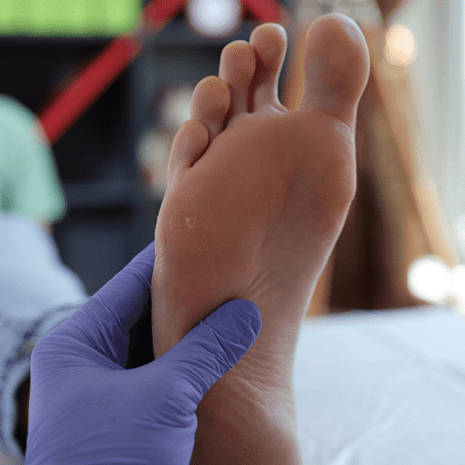
Diagnosis
Diagnosis is primarily clinical, supported by targeted tests and imaging where needed:
History & provocation: pain pattern, footwear triggers, relief with shoe removal
Physical exam: forefoot squeeze tests, palpation of webspaces, assessment for Mulder’s click
Biomechanical & gait analysis: calf length, arch profile, forefoot loading pattern
Imaging (when needed):
Ultrasound to visualise a neuroma and guide injections
MRI in complex or atypical cases, or to differentiate from intermetatarsal bursitis, stress fracture, or MTP synovitis
Diagnostic injection: local anaesthetic relief supports diagnosis
Differentials: intermetatarsal bursitis, MTP synovitis/instability, stress fracture, Freiberg disease, tarsal tunnel–related pain, capsulitis, sesamoid pathology.

Morton’s Neuroma – FAQs
It’s fibrotic thickening of a digital nerve in the forefoot due to chronic compression—not a cancer. The enlarged nerve becomes painful when squeezed between the metatarsal heads.
The third webspace (between 3rd and 4th toes) is most common; the second is next. Some patients can have multiple neuromas or bilateral symptoms.
Metatarsalgia is a general term for pain under the metatarsal heads. A neuroma specifically involves nerve thickening with burning/tingling and toe numbness, often provoked by tight shoes.
Not always. Diagnosis is typically clinical. Ultrasound is useful to confirm a lesion and guide injections; MRI helps in atypical or complex cases.
Symptoms may fluctuate, but persistent cases rarely resolve without addressing footwear, biomechanics, and load. Early conservative care improves outcomes.
A wide toe box, cushioned midsole, mild rocker, and soft forefoot uppers. Avoid narrow or high-heeled shoes that pinch the metatarsal heads together.
Yes. Metatarsal domes/pads splay the metatarsals, decompressing the nerve. Custom orthotics tailor posting and cushioning to your foot type to reduce recurrent irritation.
Ultrasound-guided corticosteroid injections can provide significant short- to medium-term relief. Alcohol sclerosing may be considered for recalcitrant cases. We guide you through risks, benefits, and expected response.
When comprehensive conservative care (orthotics, footwear, rehab ± injections) fails. Neurectomy or decompression may be offered by a surgeon; we coordinate referral and post-operative care.
Surgery can be effective, but recurrence or stump neuroma can occur. Addressing footwear and biomechanics post-op helps reduce risk.
Often yes, with shoe changes, met pads, load modification, and gait tweaks. Your plan will be individualised and progressed as symptoms settle.
Maintain appropriate footwear, continue orthotic use if prescribed, keep calf flexibility and intrinsic strength, and gradually progress training loads.
Why Choose Foot Foundation?
Foot Foundation provides specialist-level forefoot assessment with targeted offloading, orthotic design, footwear optimisation, and progressive rehabilitation. We coordinate ultrasound-guided injections and surgical referral where indicated, and support return to work and sport with graded loading plans.
With clinics in Rosedale, Takapuna, Remuera, Botany, Hamilton, and Tauranga, expert forefoot care is available across New Zealand.